
The Global Leader in Recombinant Technology:
Specialize in Antibody Development and Recombinant Protein Production
Recombinant Antibody Production Services
Sino Biological is an international reagent supplier and service provider, specializing in antibody development and recombinant protein production. We provide a variety of recombinant production and assay services for preclinical research and development.
Sino Biological has a professional mammalian cell expression platform with years of process optimization and production experience, providing one-stop services from gene synthesis to high-quality antibody production. Sino Biological has successfully delivered thousands of recombinant antibodies manufacturing programs to customers worldwide and is the preferred antibody production service provider for the world's Top10 pharmaceutical companies.
High-throughput Recombinant Antibody Production Services
100+ Abs for Screening, Delivery in 3 Weeks
| Amount | Description | Deliverables |
|---|---|---|
| 100μg |
|
|
| 500μg |
|
|
| 1mg |
|
|
Our Strengths
- HTP capabilities from gene synthesis to purified antibodies
- >10 years’ experience in antibody production
- Flexible production scales ranging from 0.05mg-5mg
- Fast delivery of purified antibodies to support screening projects
- Superior quality with high purity and low endotoxin level
- Significantly cost-effective way with very competitive price
Large-scale Recombinant Antibody Production Services
Scale-up Recombinant Antibody Production
| Amount | 10mg-100mg | 100mg-1g | >1g |
|---|---|---|---|
| 100μg | 3-5 weeks | 6-8 weeks | 8-12 weeks |
Unique Superiority
High Quality
- Monomer purity >99.9%
- Endotoxin level <0.01EU/mg
- Comprehensive QC analysis ability: ELISA, FACS, SEC-HPLC, LC-MS/MS, affinity
by OCTET…
Extensive Experience
- Various types: full mAb, chimeric, scFv, Fab, bispecific, isotype switching,
multi-chain co-transfection expression - Various species: Human, Mouse, Rat, Rabbit, Canine
- Various isotypes: IgG, IgA, IgM, IgE
Custom Antibody Development Services
Sino Biological has established high-throughput mouse & rabbit monoclonal antibody R&D platforms and also has rich experience in rabbit polyclonal antibody development.
Service Highlights:
- Free antigen evaluation & immunization scheme designing
- Speedy immunization option (mouse/rabbit): as fast as 5 weeks!
- Versatile antibody screening platform to obtain as many clones as possible that
meet custom standards - Integrated service packages (hybridoma sequencing, recombinant antibody
Workflow of Antibody Development

Project Evaluation
Expert-guided custom project design
Antigen Preparation
Peptide synthesis, recombinant protein expression

Animal Immunization
Speedy immunization could be as fast as 5 weeks
Antibody Development
Well-established hybridoma, phage display and single B cell technologies
Recombinant Antibody Production
Proprietary HEK293 platform supports high-throughput and large-scale antibody production
Fast Mouse mAb Production Service
Immunuzation time can be shortened to 5 weeks
Fast Rabbit pAb Production Service
pAb production & identification can be completed within 45 days
Rabbit mAb Production Service
Naïve human antibody library screening (phage display)

Custom Antibody Development Services
Sino Biological provides one-stop services from gene synthesis to protein expression & purification based on multiple expression systems, including bacteria, baculovirus-insect and mammalian expression systems. With more than 10 years of experience in protein expression and purification, Sino Biological has successfully expressed over 6,000 different proteins for customers worldwide.
All Recombinant Protein Production Servcices
Strengths
- Rich experience in developing >6000 protein targets
- High-efficiency expression vectors
- Proprietary transfection reagent & medium formulation
- High-density cell culture technology
- High-throughput and large-scale protein production
- One-stop service with competitive pricing and lead time
Case Studies
Recombinant Antibody production in Mammalian Cells
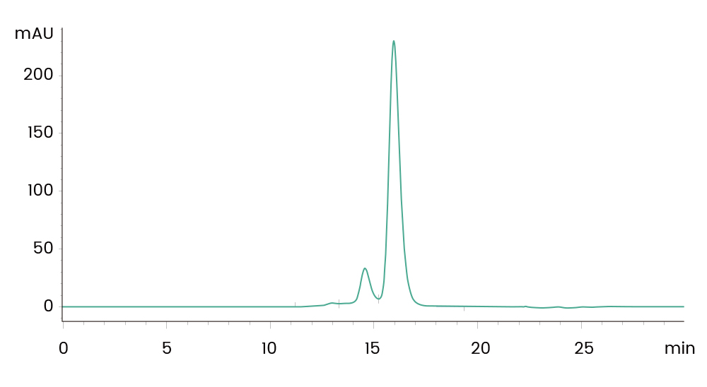
·Project:Recombinant full length antibody ·Requirements:Purity>99% by SEC-HPLC, ET ,0.5EU/mg1st Step: Protein A Affinity Chromatography
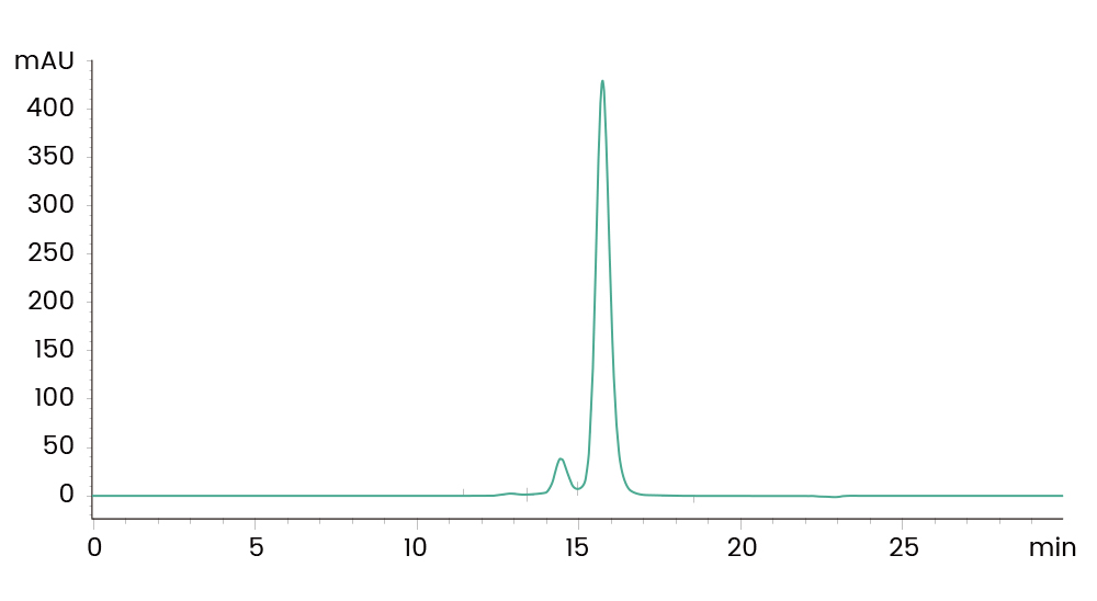
2nd step : 1st Ion Exchange Chromatography
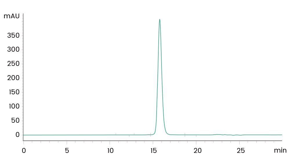
3rd step: 2nd Ion Exchange Chromatography
Custom Antibody Development
Rabbit pAb Development

Fig 1. Immunofluorescence staining of mouse target A in NIH-3T3 cells. Positive staining was localized to Mitochondrion.
Mouse mAb Development
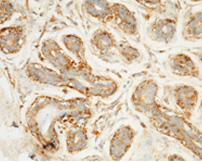
Fig 2. Immunochemical staining of human target D in human breast carcinoma with mouse mAb.
Rabbit mAb Development
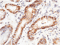
Fig 3. Immunochemical staining of human target E in human gastric cancer with rabbit mAb. The image shows membrane staining of epithelium cell.
Recombinant Antibody production in Mammalian Cells:
Tag-free Homodimer Recombinant Protein in HEK293 CellsSEC-HPLC Analysis of Target Protein after the 2nd Gel Filtrationt
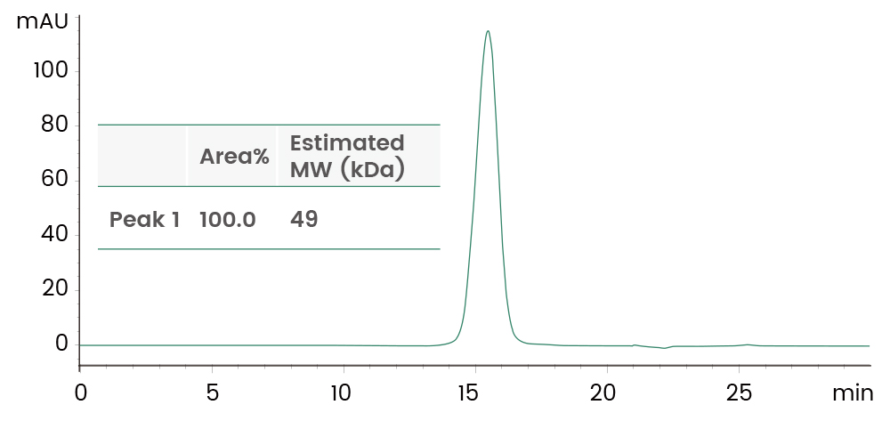
Fig 1. Immunofluorescence staining of mouse target A in NIH-3T3 cells. Positive staining was localized to Mitochondrion.
SDS-PAGE Analysis of Target Protein after the 2nd Gel Filtration
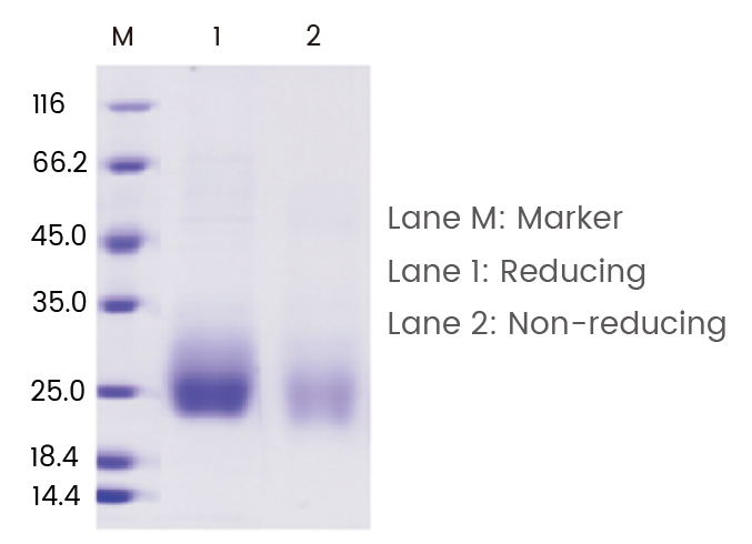
Fig 2. Immunochemical staining of human target D in human breast carcinoma with mouse mAb.
High Activity
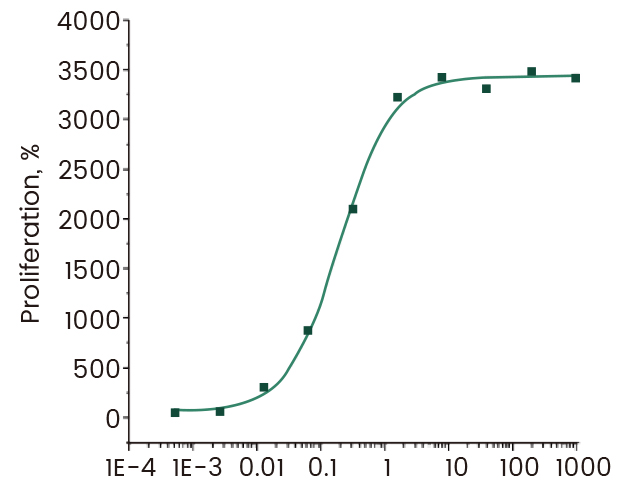
Fig 3. Immunochemical staining of human target E in human gastric cancer with rabbit mAb. The image shows membrane staining of epithelium cell.
